
Multiphoton-Based Optical Histopathology
Transforming Tissue Evaluation with Multiphoton Optical Imaging
Multiphoton-based optical histopathology is transforming tissue analysis by enabling high-resolution, label-free imaging of biological specimens without the need for traditional staining procedures.
By leveraging nonlinear optical processes such as multiphoton autofluorescence, Second Harmonic Generation (SHG), and Third Harmonic Generation (THG), researchers and clinicians can visualize tissue architecture, cellular morphology, and biochemical signatures in real time while preserving tissue integrity. This emerging approach has the potential to accelerate pathology workflows, improve diagnostic confidence, and support the transition toward digital pathology.
Based on resources from indie and Eleuthra Photonics, multiphoton imaging allows rapid, non-destructive tissue evaluation while eliminating many limitations associated with conventional staining methods.
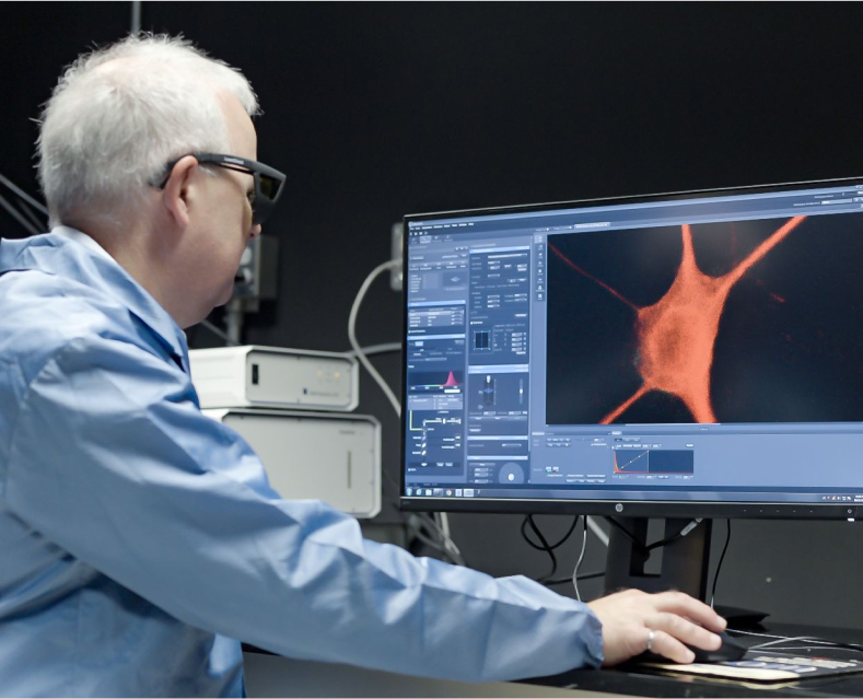
Key Applications
Digital Histopathology
Generate high-quality tissue images without staining, enabling faster pathology workflows and reducing sample preparation time.
Cancer Diagnostics
Visualize tissue morphology and tumor microenvironments with high contrast and resolution to support disease detection and characterization.
Intraoperative Tissue Assessment
Provide near real-time tissue analysis during surgical procedures, helping clinicians make informed treatment decisions.
Biomedical Research
Study tissue organization, disease progression, and cellular interactions while preserving native biological structures.
Drug Development and Translational Research
Evaluate treatment effects and tissue responses through non-destructive imaging techniques that can be repeatedly applied to samples.
Why Multiphoton-Based Histopathology Matters
Traditional histopathology relies on tissue sectioning, staining, and manual interpretation, often requiring significant preparation time and specialized laboratory workflows. Multiphoton optical histopathology offers a complementary imaging approach that can provide immediate tissue information using intrinsic biological contrast mechanisms.
Key Benefits

Label-free tissue imaging

Reduced sample preparation

Real-time visualization

High-resolution imaging

Preservation of tissue integrity

Reduced processing time

Multimodal imaging capabilities

Digital pathology compatibility
Multimodal Imaging Techniques
Multiphoton Autofluorescence
Leverages naturally occurring fluorophores within tissue to reveal metabolic and structural information without external dyes.
Second Harmonic Generation (SHG)
Provides exceptional contrast for collagen and other non-centrosymmetric structures, supporting tissue architecture analysis.
Third Harmonic Generation (THG)
Enables visualization of interfaces and tissue boundaries, complementing other nonlinear imaging modalities.
Integrated Multimodal Imaging
Combines multiple contrast mechanisms into a single platform to provide comprehensive tissue characterization.
Related Products
Femtosecond Laser – VINCI-1064

Most cost-effective fiber laser with high peak power and sub-60 fs pulse duration for high conversion efficiency.
Resources
Enhancing Real-Time Histopathology with the VINCI-1064: A Case Study Featuring Eleuthra Photonics’ TetraQuant™
Learn how Eleuthra Photonics leveraged the VINCI-1064 femtosecond fiber laser to develop an advanced multimodal nonlinear imaging system for digital histopathology.
Enhancing Multimodal Label-Free Imaging with Low-Noise VINCI-1064 fs Laser
Discover how low-noise ultrafast laser technology improves image quality and imaging performance in label-free biomedical imaging applications.
Multiphoton Microscopy with Ultrafast Fiber Lasers
Explore the fundamentals of multiphoton microscopy and its role in advanced biomedical imaging.