


Multiphoton microscopy is an advanced imaging technique that leverages the simultaneous absorption of multiple photons to excite fluorophores, generating a signal only at the focal point of the laser beam. This unique characteristic enables deeper tissue penetration, reduced out-of-focus light interference, and minimal phototoxicity, making it an indispensable tool for imaging living cells, neuronal structures, and complex biological environments. Additionally, this technique allows for real-time, high-resolution imaging of dynamic processes, making it particularly valuable in neuroscience, oncology, and regenerative medicine.
At the core of this technology are ultrafast lasers, which provide the high peak power necessary for efficient multiphoton excitation while maintaining a low average power to avoid excessive thermal damage to biological specimens. To address the growing popularity of multiphoton microscopy, there is a need for ultrafast laser sources with high peak power and excellent beam quality that are also robust, cost-effective, and compact. indie offers the VINCI, a femtosecond fiber laser specifically designed for multiphoton microscopy. It represents a significant advancement in this field, offering an optimal balance between power, precision, and cost-effectiveness. With its long emission wavelength, it extends imaging depth while minimizing scattering, ensuring enhanced signal clarity and deeper visualization of biological structures compared to traditional shorter-wavelength lasers.
Femtosecond Lasers for Multiphoton Microscopy
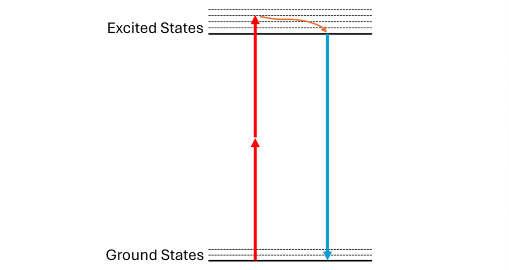
Multiphoton microscopy (MPM) encompasses several advanced techniques for imaging biological samples using nonlinear optical processes. The most common type of multiphoton microscopy is two-photon fluorescence microscopy, where two photons from the laser source are simultaneously absorbed by a fluorophore. A small amount of energy is lost as heat, and then a fluorescence signal is emitted in the form of a single photon with a higher energy (thus a shorter wavelength) (Figure 1). Other techniques, such as Second Harmonic Generation (SHG) microscopy, are energy conserving – no heat is released in the sample from the process. Techniques that rely on the simultaneous absorption of three photons are also common. All these processes allow for deeper tissue penetration and reduced photodamage compared to single-photon excitation1.
To enable effective multiphoton microscopy, a high photon density is required to achieve the nonlinear optical process. In fact, the intensity of the emitted signal is proportional to the square of the laser pulse’s peak power. Consequently, femtosecond lasers, which provide ultrashort pulses with high peak power, are essential to ensure sufficient photon flux to induce the multiphoton absorption. Since their average power remains low, these lasers also minimize thermal damage to the sample. Additionally, high peak power enhances the signal-to-noise ratio, leading to clearer and more detailed images.
Although titanium-sapphire (Ti:Sa) lasers have been the gold standard for MPM in the past due to their excellent beam quality and stability, they suffer from a few limitations. These laser systems struggle to provide sufficient power at longer wavelengths (>900 nm), which is highly desirable for deeper tissue penetration. Indeed, undesirable light scattering is inversely proportional to the fourth power of the excitation wavelength. Furthermore, Ti:Sa lasers are expensive, occupy significant space, and require regular maintenance. Their liquid cooling system also produces noticeable noise. Therefore, air-cooled fiber lasers, which are more robust and compact, are gaining popularity for MPM applications.
The VINCI series from indie
The VINCI femtosecond fiber laser has been engineered to address the growing demands of multiphoton microscopy. It integrates multiple technical features that enhance imaging performances:
- Typical pulse duration of 50 fs, optimizing multiphoton absorption efficiency.
- Wavelength >900 nm, ideal for reducing undesirable background autofluorescence and enabling deep tissue penetration by reducing scattering and absorption by biomolecules. With a 1064 nm emission wavelength, the VINCI-1064 also offers optimal compatibility with red and near-infrared fluorophores; the emission spectrum aligns with widely used biological fluorophores, maximizing excitation efficiency and signal quality.
- High Peak Power up to 1 MW for the VINCI-1064, ensuring efficient excitation even at significant imaging depths.
- Compact and Robust Design, enhancing long-term stability of the system. The VINCI series contains a simple, SESAM-free all-fiber oscillator design with a small footprint on the optical table.
- Cost-Effective Design, making high-quality multiphoton microscopy accessible at a lower overall cost compared to other ultrafast laser systems.
- Tunable Dispersion Pre-Compensation, ensuring optimal pulse duration at the biological sample by precise compensation of the chromatic dispersion of the microscope optics.
By integrating this laser into MPM systems, researchers benefit from improved image precision, higher contrast, and reduced side effects associated with prolonged irradiation. The affordability of the VINCI series makes it an attractive option for laboratories seeking high-performance imaging solutions without excessive financial investment.
Broader Applications and Future Perspectives
The application of femtosecond lasers like the VINCI to MPM extends far beyond cellular imaging, influencing multiple scientific and medical fields. These systems have become integral in a range of disciplines where high-resolution, deep-tissue imaging and minimal phototoxicity are paramount.
- Neuroscience: Multiphoton microscopy has revolutionized brain imaging, allowing researchers to visualize neural structures and activity deep within brain tissue2. This has been particularly useful in studying synaptic plasticity, neurodegenerative diseases, and real-time neuronal signaling in live specimens. Advances in femtosecond lasers have enabled functional imaging of the brain with higher precision and reduced damage to surrounding tissues.
- Oncology: Cancer research benefits significantly from femtosecond lasers, which facilitate early detection of malignant cells through non-invasive imaging techniques. These lasers enable the identification of subtle cellular changes that could indicate cancer progression, allowing for more accurate diagnostics and targeted treatments3. Additionally, multiphoton imaging is increasingly used to study tumor microenvironments and monitor therapeutic responses in real time.
- Biomaterials Research: The ability to analyze biological and synthetic materials at a microscopic level has opened new avenues in biomaterials research. Femtosecond lasers support high-resolution structural analysis, enabling researchers to examine the composition, mechanical properties, and biological interactions of various biomaterials without destructive sample preparation.
- Tissue Engineering and Regenerative Medicine: Visualizing engineered tissues at high resolution is crucial for understanding cellular organization and matrix composition in regenerative medicine. Multiphoton microscopy allows for detailed examination of bioengineered constructs, aiding in the development of advanced scaffolds, tissue grafts, and artificial organs. These imaging capabilities are instrumental in optimizing tissue regeneration strategies and assessing the integration of implants with native tissues.
- Pharmacology and Drug Discovery: In drug development, multiphoton microscopy enables high-throughput screening of pharmaceutical compounds in living tissue models. By using femtosecond lasers to track molecular interactions and drug diffusion in real-time, researchers gain deeper insights into pharmacodynamics and bioavailability, leading to more effective therapeutics4.
As ultrafast laser technology continues to advance, MPM is expected to achieve even greater imaging depth, resolution, and contrast while minimizing adverse effects on biological samples. The development of more compact, cost-effective, and power-efficient femtosecond lasers will further expand their accessibility across research and clinical fields, making this cutting-edge technology a staple in biomedical innovation.
Conclusion
Ultrafast femtosecond lasers play a central role in advancing multiphoton microscopy. Their unique characteristics enable researchers to obtain high-quality images while maintaining sample integrity. While traditional titanium-sapphire lasers have long been the gold standard, the demand for more efficient, compact, and cost-effective solutions has led to the rise of femtosecond fiber lasers. The VINCI series from indie represents a significant step forward in this field, offering a powerful yet accessible alternative for researchers seeking high-performance multiphoton imaging.
With its optimized pulse duration, high peak power, and long emission wavelength, the VINCI laser enhances imaging capabilities by minimizing scattering and maximizing fluorophore excitation efficiency. Its robust all-fiber design ensures long-term stability, while its cost-effective nature makes cutting-edge multiphoton microscopy more accessible to a broader range of laboratories. Whether in neuroscience, oncology, biomaterials research, or regenerative medicine, the VINCI laser is pushing the boundaries of deep-tissue imaging and biomedical discovery.
As multiphoton microscopy continues to evolve, the integration of next-generation femtosecond lasers like the VINCI will drive new breakthroughs in biological and medical research, making high-resolution, real-time imaging an indispensable tool for scientists and clinicians alike.
- Larson A., Multiphoton microscopy. Nature Photon, 2011, vol. 5, no. 1 (2011). ↩︎
- Lecoq J, Orlova N, Grewe B. F., Wide. Fast. Deep: Recent Advances in Multiphoton Microscopy of In Vivo Neuronal Activity. J. Neurosci, 2019, vol. 39, no. 46, pp. 9042-9052 ↩︎
- Ávila F. J., A Review of Non-Linear Optical Imaging Techniques for Cancer Detection. Optics, 2024, vol. 5, no. 4, pp. 416-433 ↩︎
- Morimoto A, Kikuta J, Ishii M., Intravital multiphoton microscopy as a novel tool in the field of immunopharmacology. Pharmacol. Ther., 2020, vol. 206, p. 107429 ↩︎
Related Product
| Product | |
|---|---|
| Femtosecond Laser – VINCI-1064 | View Product |